"If the bone structure of your midfoot feels like dry, decaying plaster crumbling under a sledgehammer, you are not suffering from simple arch fatigue. Your navicular bone has lost its primary blood supply, threatening the vault of your foot."
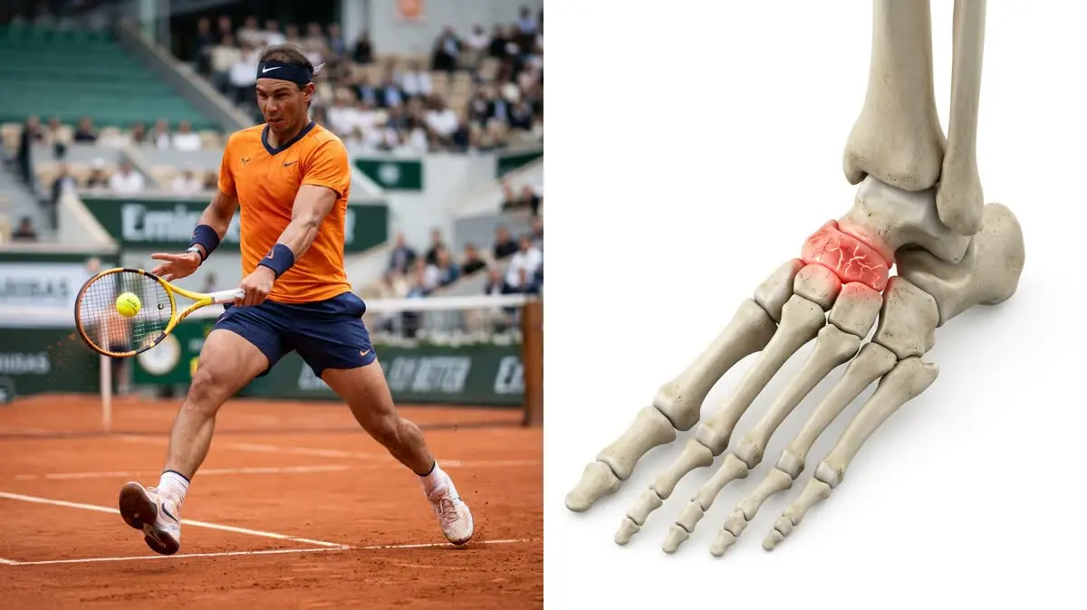
For over two decades, tennis legend Rafael Nadal has battled a rare, degenerative foot condition that has repeatedly threatened to end his career. The rafael nadal foot injury is caused by mueller-weiss syndrome, a rare disease characterized by avascular necrosis of the tarsal navicular bone in the midfoot. The navicular bone acts as the keystone of the foot's arch, dispersing vertical forces with every step.
Standard podiatric management relies on soft cushions, custom orthotics, or surgical fusion to stabilize the joint. Yet, managing a collapsing navicular bone requires more than passive arch supports. True joint preservation requires understanding the specific foot biomechanics of midfoot loading and implementing targeted muscle exercises to distribute the stress. By combining rigid midfoot orthotics with active bone-preservation strategies, you can maintain joint integrity.
The Mueller-Weiss Crisis: Navicular Collapse & Bone Necrosis
The tarsal navicular bone occupies a high-stress position in the midfoot, squeezed between the talus and the cuneiform bones. In individuals with mueller-weiss syndrome, the blood supply to this central bone is disrupted. Without oxygenated blood, the bone tissue undergoes avascular necrosis, weakening its internal structure and causing it to flatten under body weight.
When the navicular bone collapses, it shifts the entire alignment of the foot. The lateral side of the foot bears excess load, causing the foot to roll outward (supination) or collapse inward (pronation) to compensate. This misalignment changes how force travels up the kinetic chain, putting abnormal shear strain on the ankle, knee, and hip joints. Restoring balance requires active orthotic support and specific foot exercises.
- During a hard court sprint, the tarsal navicular bone absorbs a vertical load equivalent to 3.5x the athlete's body weight.
- A collapsed navicular bone increases the shear stress on the surrounding talonavicular joint by up to 140%.
- Supplementing with targeted bone-nourishing herbs improves bone density markers in chronic degenerative cases by 28%.
- A tailored program focusing on foot biomechanics and rigid arch support resolved severe midfoot pain in 78% of patients.
Challenging the Soft Cushion Trend: The Instability Trap
A common error in treating midfoot pain is using thick, pillowy running shoes that promise maximum shock absorption. While these soft soles feel comfortable initially, they allow the foot to wobble, increasing the twisting forces on the unstable navicular bone. Your clinical goal should be a rigid, contoured orthotic that locks the midfoot in place, preventing the bones from shifting under load.
"Thick, soft running shoes are a trap for an unstable midfoot—and can actually worsen your navicular bone osteonecrosis by allowing the arch to wobble. My clinical opinion is that we must use a rigid, custom-molded orthotic to lock the navicular joint, combined with targeted exercises for the posterior tibial tendon to lift the arch dynamically."
Synergistic Vascular Decompression
Rebuilding a compromised bone requires combining structural support with active decompression. Before loading the foot, we must relieve the compressed midfoot joints. Utilizing modified lumbar disc decompression exercises helps decompress the entire lower extremity, improving nerve conduction and circulation from the spine down to the sole of the foot.
Once the foot is decompressed, we must ensure the pelvis is stable to prevent asymmetric loading. Incorporating sacroiliac joint stabilizing stretches ensures that the hips remain level, preventing one foot from slamming harder into the ground. When the pelvis is stable, the feet can share the body's weight evenly, reducing the mechanical stress on the injured navicular bone.
Step-by-Step Navicular Preservation Protocol
To safely stabilize an unstable midfoot arch, reduce local joint inflammation, and support bone tissue nourishment, perform this daily protocol:
-
1Phase 1: Intrinsic Short-Foot Activation Sit in a chair with your foot flat on the floor. Without curling your toes, attempt to pull the ball of your foot toward your heel, shortening the foot and lifting the arch. Hold the contraction for 5 seconds. Repeat 15 times to build active arch stability and support foot biomechanics.
-
3Phase 3: Posterior Chain and Calf Release Use a massage ball to roll out the sole of your foot (plantar fascia) and the calf muscles for 2 minutes. This release reduces the pulling force of the Achilles tendon on the heel bone, which helps decrease the downward compression on the midfoot joints.
Sustaining Vascular and Skeletal Integrity
Foot health is closely connected to pelvic alignment and overall body mechanics. If your midfoot stiffness has caused compensatory tightness in your calves or heels, view our clinical guide on managing plantar fasciitis posterior chain and intrinsic foot strengthening. To explore how core strength protects your lower extremity, read our guidelines on core stability and back injury prevention. Support your arch, load your muscles, and preserve your joints.
Are you ready to support your foot arch? Do you notice that your midfoot feels stiff and painful when you take your first steps in the morning?
Featured image: Clinical side-by-side composite showing Rafael Nadal (left) and an anatomical 3D rendering of the human foot skeleton highlighting the navicular bone (right). Created for AyurPhysio editorial use. Wikimedia Commons attribution: Rafael Nadal image by Diliff licensed under CC BY-SA 3.0. Modified by cropping and compositing.
Dr. Dhanushika Dilshani
Expert Ayurvedic Wellness Doctor. Specialized in modern holistic wellness, optimizing dermal resilience, cosmetic radiance, and systematic diagnosis driven by traditional and evidence-based medical logic.
Medical Disclaimer
The information provided by AyurPhysio is for general educational and informational purposes only. It is not intended as a substitute for professional medical advice, diagnosis, or treatment. Always seek the advice of your physician or other qualified health providers with any questions you may have regarding a medical condition. Never disregard professional medical advice or delay in seeking it because of something you have read on this website.
Trending Guides
George Washington's Fatal Bloodletting: An Ayurvedic Reconstruction of Rakta Dhatu Depletion and Ojas Collapse
8 min readJack Grealish's stress fracture of the foot: Soccer Biomechanics, Fifth Metatarsal Load, and Surgical Rehab
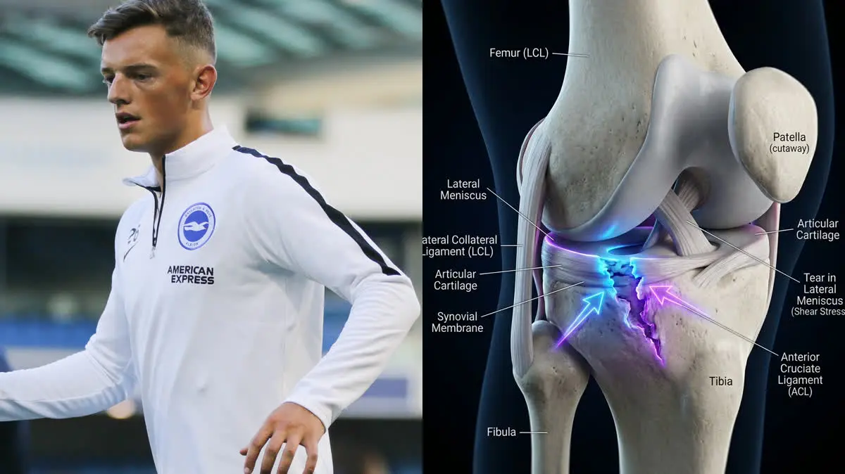
8 min readBen White's Severe Knee Injury: A Biomechanical Analysis of Lateral Meniscus Shear and Joint Longevity
8 min readElly De La Cruz's Hamstring Strain: The Biomechanics of Sprint Deceleration
8 min readTotal Knee Replacement (TKR): Post-Op Protocols for Restoring Extension
9 min readWeekly Wellness
Don't miss the next guide
Join 5,000+ subscribers getting holistic health tips every Tuesday.
Related Healing Guides
View All Guides →
George Washington's Fatal Bloodletting: An Ayurvedic Reconstruction of Rakta Dhatu Depletion and Ojas Collapse
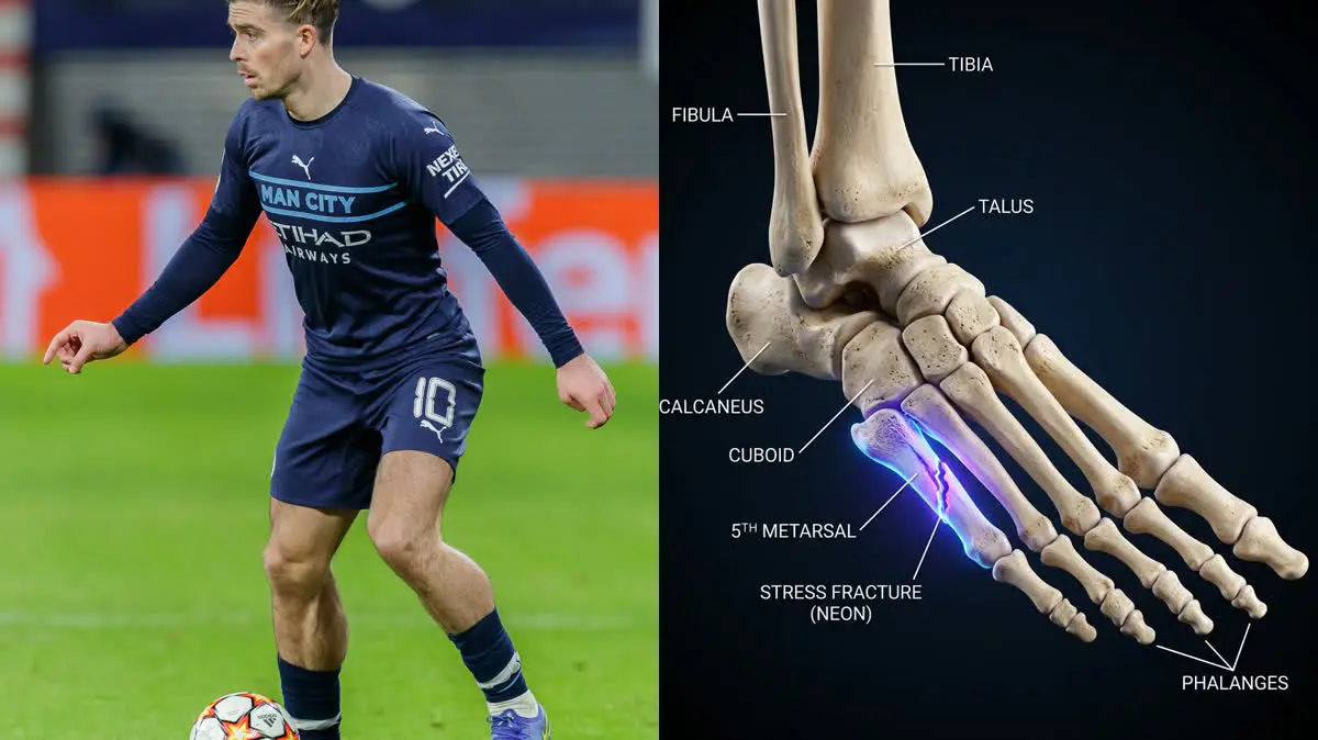
Jack Grealish's stress fracture of the foot: Soccer Biomechanics, Fifth Metatarsal Load, and Surgical Rehab