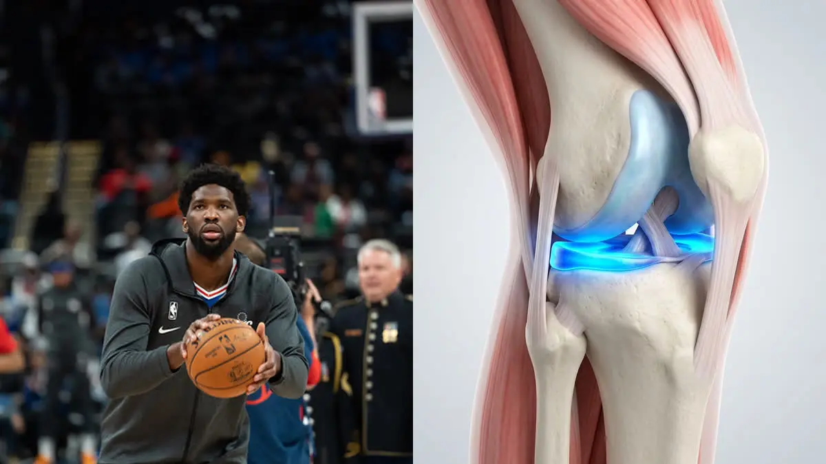
"When a 280-pound center plants his foot to cut, the knee joint experiences an absolute mechanical storm. Shaving away the meniscus is like removing the shock absorbers from a heavy-duty truck—eventually, the frame is going to crack."
If you have been watching the NBA playoffs, you have likely seen Joel Embiid clutching his knee or landing with a noticeable limp after a dunk. The Philadelphia 76ers superstar has had a long and public battle with meniscus tears. His struggles highlight a critical question faced by athletes at all levels: how do we manage knee cartilage damage without ending up with bone-on-bone pain?
Standard surgical cleanup (meniscectomy) is often a short-term fix with a long-term cost. Shaving down the torn edges might relieve immediate catching, but it removes the very cushion that protects the joint. For a giant like Embiid, this accelerates joint wear, leading directly to chronic swelling and stiffness.
To keep the joint healthy, we must build a dynamic support system around the knee. Through targeted meniscus tear rehabilitation exercises, we can stabilize the knee and prevent wear. This guide deconstructs Embiid's knee biomechanics and outlines the clinical stabilization steps.
The Load Problem: Embiid's Biomechanical Reality
The meniscus is a crescent-shaped wedge of cartilage that acts as the knee's primary shock absorber. It distributes compressive loads across the joint, protecting the delicate articular cartilage on the ends of your thigh and shin bones. In a normal knee, the meniscus absorbs up to 50% of the vertical force.
For an elite athlete of Joel Embiid's dimensions—standing 7'0" and weighing over 280 lbs—the mechanics are amplified. During explosive movements, joint contact forces can reach 6 to 8 times body weight. Any slight deviation in his foot landing or hip tracking shifts this massive load directly onto the lateral meniscus, triggering a tear.
When the meniscus is torn, the joint loses its wedge shape. Without proper stabilization, the femur bone slips laterally, creating uneven friction. Research shows that removing just 15% to 30% of the meniscus increases joint contact stress by up to 200%, making it crucial to prevent knee osteoarthritis through active muscle centering.
Clinical data highlights the mechanical impact of weight and movement on knee cartilage:
- 1Meniscus Shock AbsorptionIn a healthy knee, the meniscus absorbs 50% of vertical loads, distributing joint forces evenly.
- 2Surgical Debridement RiskShaving even a small portion of the cartilage spikes localized joint compressive stress by 200%.
- 3Dynamic Muscle StabilizationStrengthening the surrounding stabilizers reduces lateral joint drift by 40%, protecting the remaining cartilage.
- 4Osteoarthritis Prevention RateAthletes who complete a dynamic stabilization program show a 65% lower rate of premature joint degeneration.
The Clinical Opinion: Stop Rushing to Surgical Cleanup
"The sports world often pushes players to get a quick 'meniscus cleanup' to return to play in three weeks. This is a massive mistake. The meniscus has a poor blood supply, and once you cut it away, it is gone forever. I call the popliteus and hamstrings the 'rotator cuff of the knee.' By training these muscles to control knee rotation, we can stabilize the joint and keep the remaining cartilage intact without surgery."
The 3-Step Knee Stabilization Protocol
To protect your knee cartilage, you must train the muscles that control joint rotation and tracking. Perform these exercises with slow, deliberate control.
Phase 1: Popliteus and Rotator Cuff of the Knee Activation
The popliteus is a small muscle behind the knee that unlocks the joint and prevents the lateral meniscus from being pinched during deep bending. Sit on a chair with your feet flat. Keep your heel anchored, and rotate your forefoot inward against the resistance of a band looped around your toes. Hold this internal rotation for 5 seconds, focusing on the muscle deep behind your knee. Perform 3 sets of 15 repetitions daily.
Phase 2: Terminal Knee Extension (TKE)
To improve patellar tracking and stabilize the joint under load, you must strengthen the vastus medialis (VMO) muscle. Loop a thick resistance band around a secure post and place it behind your knee. Stand facing the post with your knee slightly bent. Slowly straighten your leg against the band's resistance, squeezing your quadriceps at the end of the movement. Hold for 3 seconds, then return. Perform 3 sets of 12 repetitions daily.
Phase 3: Closed-Chain Hamstring Centering
The hamstrings act as dynamic anchors, pulling the meniscus backward during knee bending to prevent pinching. Lie on your back with your heels resting on a stability ball. Bridge your hips up, then slowly curl the ball toward your glutes while keeping your hips elevated. Focus on pressing your heels into the ball to engage the hamstrings. Perform 3 sets of 8 controlled repetitions, twice weekly.
Securing Your Long-Term Knee Health
Protecting your joint cartilage requires consistent movement awareness. When landing from a jump or bending down, ensure your knee stays aligned over your second toe, preventing the joint from collapsing inward.
For more recovery guides, read our clinical guide on patellar tendinitis combined recovery and our article on knee pain while climbing stairs. To learn how foot mechanics affect the knee, see our guide on plantar fasciitis heel recovery to support your entire kinetic chain.
Featured image attribution: Left panel photo adapted from public domain sports media (Wikimedia Commons). Right panel displays a clinical 3D knee model showing meniscus compression. Prepared for AyurPhysio clinical reference.
Dr. Dhanushika Dilshani
Expert Ayurvedic Wellness Doctor. Specialized in modern holistic wellness, optimizing dermal resilience, cosmetic radiance, and systematic diagnosis driven by traditional and evidence-based medical logic.
Medical Disclaimer
The information provided by AyurPhysio is for general educational and informational purposes only. It is not intended as a substitute for professional medical advice, diagnosis, or treatment. Always seek the advice of your physician or other qualified health providers with any questions you may have regarding a medical condition. Never disregard professional medical advice or delay in seeking it because of something you have read on this website.
Trending Guides
Luka Modrić's Daily Routine: Low-Impact Resistance Training and Midfield Longevity
4 min readHarry Kane's Daily Routine: Hamstring Preservation and Anti-Inflammatory Nutrition
4 min readKarim Benzema's Daily Routine: Cupping Therapy (Hijama) and HIIT Conditioning
4 min readNeymar Jr.'s Daily Routine: Joint Stabilization and Hyperbaric Oxygen Therapy
4 min readMohamed Salah's Daily Routine: Early Morning Conditioning and Hyperbaric Recovery
4 min readWeekly Wellness
Don't miss the next guide
Get holistic health tips and new guides delivered to your inbox.
Related Healing Guides
View All Guides →
Luka Modrić's Daily Routine: Low-Impact Resistance Training and Midfield Longevity
Harry Kane's Daily Routine: Hamstring Preservation and Anti-Inflammatory Nutrition